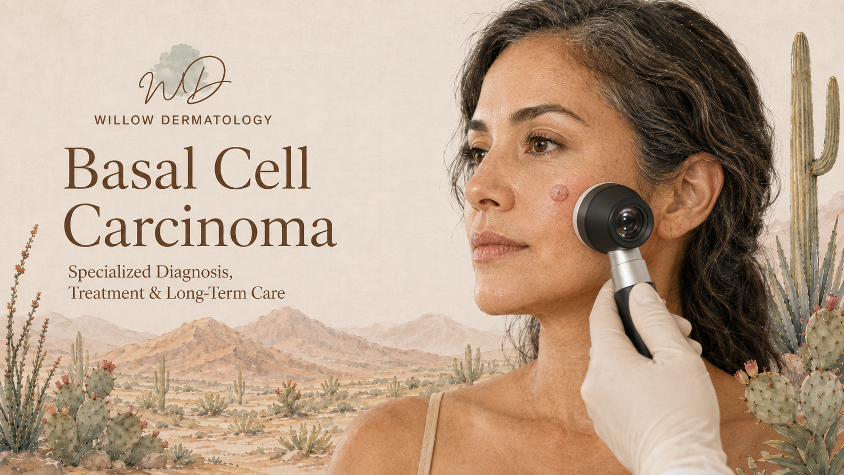

Dermatologists Treating Basal Cell Carcinoma in Tucson
What Is Basal Cell Carcinoma?
Basal cell carcinoma is the most common type of skin cancer. It develops from basal cells located in the outermost layer of the skin and most often appears on areas that have received significant sun exposure, particularly the face, ears, scalp, neck, shoulders, and arms.
Basal cell carcinoma usually grows slowly and rarely spreads to distant areas of the body. However, it should not be ignored. Without treatment, a basal cell carcinoma can continue growing into nearby skin, cartilage, nerves, muscle, or bone. Early diagnosis generally allows for simpler treatment and helps preserve as much healthy tissue as possible.
What Does Basal Cell Carcinoma Look Like?
Basal cell carcinoma can have several different appearances. It may look like:
A pearly, shiny, or translucent bump
A pink, red, brown, black, or skin-colored growth
A scaly patch that does not resolve
A sore that repeatedly bleeds, crusts, heals, and returns
A scar-like area without a history of injury
A slowly enlarging spot with rolled or raised borders
Basal cell carcinoma can occur in people of every skin tone. Any new, changing, bleeding, or nonhealing lesion should be evaluated by a dermatologist.
Diagnosis and Risk Assessment
Diagnosis usually begins with a careful examination followed by a skin biopsy. The biopsy confirms the diagnosis and identifies the microscopic growth pattern of the tumor.
Basal cell carcinomas are not all the same. Common subtypes include nodular, superficial, infiltrative, micronodular, and morpheaform basal cell carcinoma. Some subtypes grow in narrow strands beneath the skin and may extend farther than the visible surface lesion suggests.
Treatment planning considers the tumor’s size, location, borders, microscopic subtype, whether it has been treated previously, and the patient’s overall health and immune status. Tumors involving the face, ears, scalp, hands, feet, or other functionally important areas may require a more specialized approach.
Treatment Options
Treatment is individualized to completely remove or destroy the cancer while preserving healthy tissue and achieving the best possible functional and cosmetic result.
Mohs micrographic surgery is often recommended for basal cell carcinomas that are located on the face or other tissue-sensitive areas, have an aggressive microscopic pattern, are recurrent, have poorly defined borders, or otherwise carry a higher risk of returning. During Mohs surgery, the tumor is removed in stages and the complete surgical margin is examined microscopically. This allows the surgeon to remove the cancer while conserving as much unaffected tissue as possible.
Standard surgical excision may be appropriate for many well-defined tumors. The cancer and a measured margin of surrounding skin are removed and sent to a laboratory for examination.
Curettage and electrodesiccation may be used for selected low-risk tumors in appropriate locations. The cancerous tissue is removed with a specialized instrument, and the base is treated with controlled electrical energy.
Topical medications, including imiquimod or fluorouracil, may be considered for certain superficial basal cell carcinomas. These treatments are not appropriate for every tumor and require careful selection and follow-up.
Radiation therapy or systemic medication may be considered when surgery is not appropriate or when a tumor is locally advanced, recurrent, or has rarely spread beyond its original location. Complex cases may be managed collaboratively with radiation oncology or medical oncology.
Follow-Up and Prevention
Having one basal cell carcinoma increases the likelihood of developing another skin cancer in the future. Continued dermatologic surveillance is therefore an important part of care.
Follow-up recommendations are individualized based on your history and risk factors. Regular skin examinations, monthly self-checks, protective clothing, broad-spectrum sunscreen, and avoidance of tanning beds can help identify future skin cancers earlier and reduce additional ultraviolet damage.
If you notice a new growth, a changing spot, or a sore that repeatedly bleeds or does not heal, schedule a dermatologic evaluation.