
Dermatologists Treating Cutaneous Lymphomas in Tucson
Cutaneous Lymphomas
Willow Dermatology provides specialized evaluation, diagnosis, treatment coordination, and long-term surveillance for cutaneous lymphomas, including cutaneous T-cell lymphoma, its variants, and primary cutaneous B-cell lymphoma. Care is individualized according to the lymphoma subtype, extent of skin involvement, symptoms, microscopic findings, and whether the disease involves the blood, lymph nodes, or internal organs.
What Is Cutaneous Lymphoma?
Cutaneous lymphomas are uncommon cancers of lymphocytes, the white blood cells that help regulate the immune system. When a lymphoma begins primarily within the skin, it is called a primary cutaneous lymphoma.
These cancers are different from lymphomas that begin in the lymph nodes or another internal organ and later spread to the skin. Determining whether a lymphoma is primary to the skin or part of a systemic lymphoma is an essential part of diagnosis and treatment planning.
Cutaneous lymphomas are broadly divided into:
Cutaneous T-cell lymphomas, which develop from T lymphocytes
Cutaneous B-cell lymphomas, which develop from B lymphocytes
The clinical course varies considerably. Some cutaneous lymphomas remain limited to the skin and progress slowly over many years, while others require prompt multidisciplinary treatment because they have a greater potential to involve the blood, lymph nodes, or internal organs.
What Does Cutaneous Lymphoma Look Like?
Cutaneous lymphoma can resemble eczema, psoriasis, dermatitis, an insect bite, a cyst, or another common skin condition. Possible signs include:
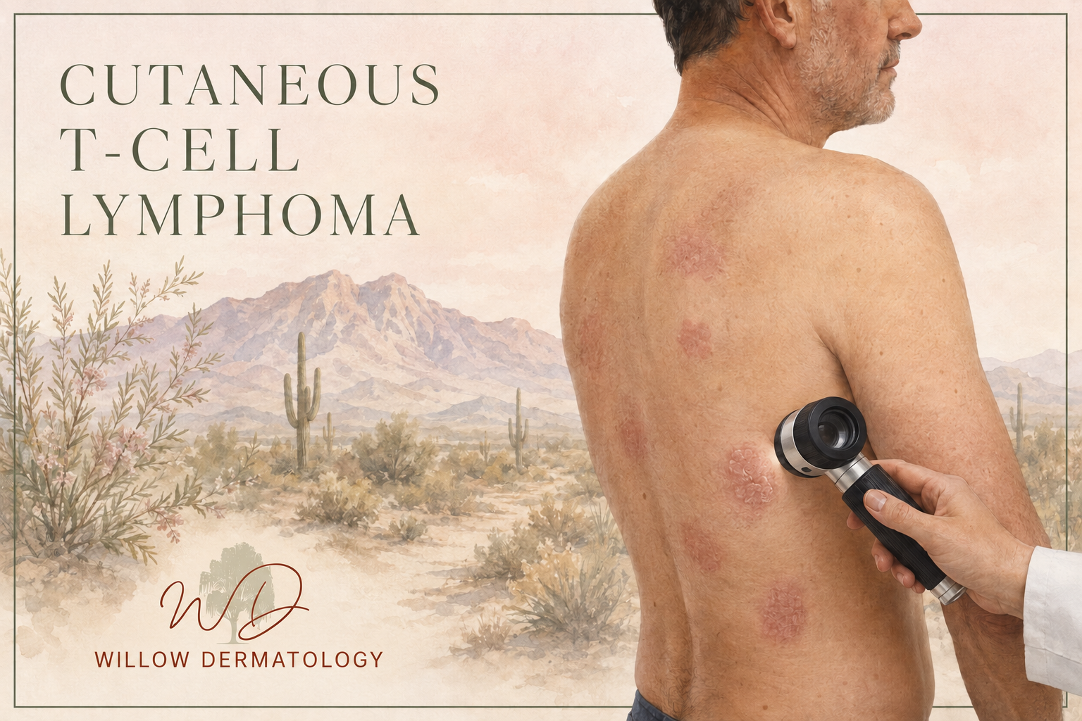
Persistent dry, scaly, or discolored patches
Areas of skin that are repeatedly itchy, irritated, or painful
Thickened or raised plaques
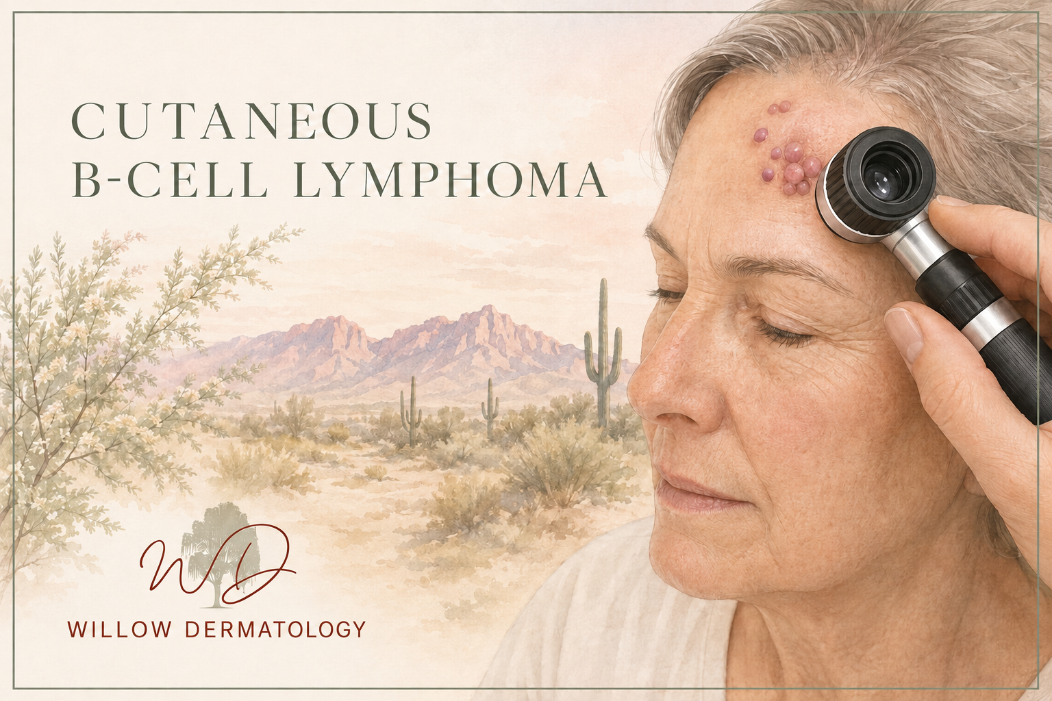
Smooth pink, red, or violet bumps
Firm nodules beneath or within the skin
Crops of bumps that appear, crust, and heal
Tumors that enlarge or develop surface breakdown
Widespread redness, scaling, or severe itching
Unexplained hair loss within an affected area
Enlarged lymph nodes or swelling beneath the skin
Because early cutaneous lymphoma can closely resemble an inflammatory rash, the diagnosis may require more than one biopsy and careful comparison of the clinical and microscopic findings.
Cutaneous T-Cell Lymphoma
Cutaneous T-cell lymphoma is a group of lymphomas in which abnormal T lymphocytes primarily involve the skin. The most common type is mycosis fungoides, but several additional variants and related disorders can produce different symptoms and follow different clinical courses.
Mycosis Fungoides
Mycosis fungoides is the most common form of cutaneous T-cell lymphoma. It often begins with flat, scaly, or discolored patches that may resemble eczema or psoriasis. These areas commonly develop on the trunk, buttocks, hips, or other locations that receive limited sun exposure.
In some patients, the disease remains in the patch stage for many years. In others, patches may gradually become thicker plaques or develop into raised tumors. Itching can range from mild to severe.
Early-stage mycosis fungoides is frequently managed with treatments directed primarily at the skin. More extensive disease may require systemic therapy and coordinated care with hematology-oncology or radiation oncology.
Variants of Mycosis Fungoides
Several less common forms of mycosis fungoides have distinctive clinical features:
Folliculotropic mycosis fungoides involves the hair follicles and commonly affects the head and neck. It may cause grouped bumps, acne-like lesions, thickened plaques, itching, or localized hair loss. Because the abnormal cells may extend more deeply around hair follicles, treatment may differ from that used for superficial patch-stage disease.
Pagetoid reticulosis usually appears as a single, slowly enlarging scaly patch or plaque. It most often remains localized to one area of skin and generally has a favorable clinical course.
Granulomatous slack skin is an extremely rare variant that can cause loose or folded areas of skin, particularly within body folds. Diagnosis requires correlation between the physical examination and specialized microscopic findings.
Sézary Syndrome
Sézary syndrome is a less common but more advanced form of cutaneous T-cell lymphoma involving the skin, blood, and usually the lymph nodes.
Patients may develop widespread redness and scaling over most of the body, severe itching, swelling, thickening of the palms or soles, hair loss, nail changes, or enlarged lymph nodes. Blood testing and flow cytometry are important for identifying abnormal circulating T lymphocytes and establishing the diagnosis.
Sézary syndrome generally requires systemic treatment and coordinated management involving dermatology, hematology-oncology, and other specialists.
Primary Cutaneous CD30-Positive Lymphoproliferative Disorders
This group includes lymphomatoid papulosis and primary cutaneous anaplastic large cell lymphoma.
Lymphomatoid papulosis causes recurring crops of red or violet bumps that may crust, break down, and then heal on their own. Although the individual lesions often resolve, the abnormal cells have lymphoma-like features under the microscope. Patients require long-term surveillance because they have an increased risk of developing another lymphoid malignancy.
Primary cutaneous anaplastic large cell lymphoma usually appears as one or more rapidly enlarging red or violet nodules or tumors. Some lesions may develop crusting or ulceration. The disease often remains limited to the skin, but appropriate staging is necessary to exclude lymph node or internal involvement.
Other Rare Cutaneous T-Cell Lymphomas
Additional uncommon disorders include:
Primary cutaneous CD4-positive small or medium T-cell lymphoproliferative disorder
Subcutaneous panniculitis-like T-cell lymphoma
Primary cutaneous gamma-delta T-cell lymphoma
Primary cutaneous aggressive epidermotropic cytotoxic CD8-positive T-cell lymphoma
Other rare peripheral T-cell lymphomas involving the skin
Some of these conditions follow a relatively slow and localized course, while others may progress more rapidly. Accurate classification by a dermatopathologist with expertise in cutaneous lymphoma is essential because treatment and prognosis differ substantially among these diagnoses.
Primary Cutaneous B-Cell Lymphoma
Primary cutaneous B-cell lymphomas begin from abnormal B lymphocytes within the skin. They most often appear as smooth, firm, pink, red, or violet papules, plaques, or nodules.
The three principal subtypes have different patterns of behavior and treatment.
Primary Cutaneous Follicle Center Lymphoma
Primary cutaneous follicle center lymphoma is an indolent lymphoma that most commonly develops on the scalp, forehead, or trunk. It may appear as a solitary growth or a cluster of firm, smooth papules, plaques, or nodules.
This lymphoma frequently remains limited to the skin and generally has a favorable prognosis. Localized disease may be treated with radiation therapy or surgical excision. Patients with multiple lesions may require additional skin-directed or systemic therapy.
Primary Cutaneous Marginal Zone B-Cell Lymphoma
Primary cutaneous marginal zone B-cell lymphoma, also described as a primary cutaneous marginal zone lymphoproliferative disorder in some classifications, is usually slow-growing and confined to the skin.
It commonly produces one or more red to violet bumps or plaques on the trunk or arms. Treatment may include observation in selected cases, radiation therapy, surgical excision, injected medication, or systemic therapy, depending on the number, location, and behavior of the lesions.
Primary Cutaneous Diffuse Large B-Cell Lymphoma, Leg Type
Primary cutaneous diffuse large B-cell lymphoma, leg type, is a more aggressive form of cutaneous B-cell lymphoma. It most often appears as rapidly enlarging red, blue-red, or violet nodules or tumors on one or both lower legs, although it can occur elsewhere.
Prompt staging and treatment are important because this subtype has a greater risk of spreading beyond the skin. Management commonly involves systemic immunotherapy or immunochemotherapy, often combined with radiation therapy.
Diagnosis and Staging
Diagnosis begins with a detailed examination of the skin and lymph nodes followed by one or more skin biopsies. A generous biopsy may be necessary because the distribution and depth of abnormal lymphocytes provide important diagnostic information.
Specialized testing may include:
Immunohistochemical staining to identify the type of lymphocyte involved
Molecular testing for T-cell or B-cell clonality
Complete blood count and other laboratory studies
Peripheral blood flow cytometry
Review by a dermatopathologist or hematopathologist
Biopsy of an enlarged or suspicious lymph node
Computed tomography or positron emission tomography imaging
Bone marrow evaluation in selected cases
Early mycosis fungoides may be difficult to confirm with a single biopsy. When clinical concern remains despite an inconclusive result, additional biopsies obtained over time may help establish the diagnosis.
Staging is tailored to the specific lymphoma subtype. It may evaluate the amount and type of skin involvement, lymph nodes, blood, and internal organs.
Treatment Options
Treatment is individualized according to the exact diagnosis, stage, symptoms, rate of progression, and overall health of the patient.
Skin-directed treatments may be appropriate when disease is limited to the skin. Options can include topical corticosteroids, topical mechlorethamine, phototherapy, localized radiation therapy, or total skin electron beam therapy.
Surgical excision may be considered for selected solitary cutaneous B-cell lymphomas, localized primary cutaneous anaplastic large cell lymphoma, or other isolated lesions when surgery is expected to provide effective local control.
Systemic treatments may include oral retinoid medications, interferon, extracorporeal photopheresis, targeted medications, monoclonal antibody therapy, immunotherapy, or chemotherapy.
Radiation therapy can be highly effective for individual plaques, tumors, or localized cutaneous B-cell lymphomas. More extensive skin involvement may occasionally be treated with total skin electron beam therapy.
Hematopoietic stem cell transplantation may be considered for selected patients with advanced, aggressive, or treatment-resistant cutaneous lymphoma.
Because these conditions differ substantially, a treatment that is appropriate for one subtype may not be suitable for another. Complex or advanced cases are often managed collaboratively with hematology-oncology, radiation oncology, dermatopathology, and other specialists.
Follow-Up and Long-Term Surveillance
Long-term follow-up is important even when the lymphoma is slow-growing or responds completely to treatment. Cutaneous lymphomas may recur in previously treated skin, appear in new areas, or, in some cases, progress beyond the skin.
Surveillance is individualized and may include:
Regular examinations of the skin and lymph nodes
Monitoring for new patches, plaques, nodules, or tumors
Repeat laboratory testing or blood flow cytometry when indicated
Imaging for selected lymphoma subtypes or stages
Ongoing management of itching, skin dryness, and treatment-related effects
Coordination with hematology-oncology or radiation oncology
Contact your dermatologist if you develop a persistent unexplained rash, recurrent crops of bumps, a rapidly enlarging nodule, widespread redness and itching, enlarged lymph nodes, unexplained fevers, drenching night sweats, or unintentional weight loss.
Early evaluation and accurate classification allow treatment to be matched to the biological behavior of the lymphoma while avoiding unnecessary therapy for slower-growing forms.