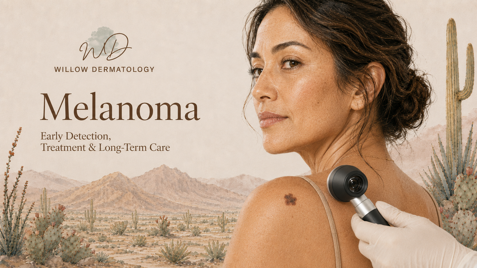

Dermatologists Treating Melanoma in Tucson
What Is Melanoma?
Melanoma is a type of skin cancer that develops from melanocytes, the cells that produce pigment in the skin. It may begin within an existing mole or appear as a new spot. Although melanoma is less common than basal cell carcinoma and squamous cell carcinoma, it has a greater potential to spread to lymph nodes and other organs.
Melanoma can develop anywhere on the body, including areas that receive little sun exposure. It may also occur on the palms, soles, beneath the nails, and, less commonly, within the eyes or mucosal surfaces. Melanoma can affect people of every age and skin tone.
When melanoma is identified and treated early, it is often highly curable. Prompt evaluation of suspicious or changing lesions is essential.
What Does Melanoma Look Like?
The ABCDE warning signs can help identify a suspicious mole or pigmented lesion:
Asymmetry: One half does not match the other
Border: The edges are irregular, blurred, scalloped, or poorly defined
Color: The lesion contains multiple shades of brown, black, tan, red, blue, or white
Diameter: The lesion is often larger than 6 millimeters, although melanomas can be smaller
Evolving: The spot changes in size, shape, color, elevation, sensation, or symptoms
Other warning signs may include:
A new mole or pigmented spot that looks different from your other moles
A lesion that grows, changes, itches, becomes painful, or begins bleeding
A pink, red, or skin-colored growth without obvious dark pigment
A dark streak beneath a fingernail or toenail
A changing spot on the palm or sole
A sore that does not heal
The “ugly duckling” sign describes a mole or spot that looks noticeably different from the others on your skin. Any new, changing, or unusual lesion should be evaluated by a dermatologist.
Diagnosis and Risk Assessment
Diagnosis begins with a careful examination of the skin, often using a dermatoscope to evaluate structures and colors that are not visible to the unaided eye. If a lesion is suspicious, a skin biopsy is performed. When possible, the entire visible lesion is removed during the initial biopsy so it can be examined accurately.
The pathology report confirms the diagnosis and provides information needed for treatment planning. Important features may include the melanoma subtype, tumor thickness, ulceration, mitotic activity, and whether the biopsy edges contain melanoma cells.
Melanoma staging considers how deeply the tumor has grown, whether it has ulcerated, and whether it has reached nearby lymph nodes or other parts of the body. Depending on the tumor’s thickness and other risk factors, additional evaluation may include lymph node examination, sentinel lymph node biopsy, laboratory testing, or imaging.
Treatment Options
Treatment is individualized according to the melanoma’s stage, location, microscopic features, and the patient’s overall health.
Wide local excision is the standard treatment for most localized melanomas. The melanoma and a measured margin of surrounding skin are surgically removed. The recommended margin is determined by the tumor’s depth and whether the melanoma is invasive or confined to the outermost layer of the skin.
Mohs micrographic surgery or staged excision may be considered for selected melanomas in situ, including lentigo maligna, particularly on the face or other anatomically sensitive areas. These techniques allow careful examination of the peripheral margins while preserving healthy tissue. Specialized immunostains may be used to help identify melanoma cells during margin assessment.
Sentinel lymph node biopsy may be recommended for selected invasive melanomas with a meaningful risk of spreading. This procedure examines the first lymph node or group of lymph nodes most likely to receive cancer cells from the original tumor.
Immunotherapy, targeted therapy, radiation therapy, or additional surgery may be recommended for higher-risk or advanced melanoma. These cases are managed collaboratively with surgical oncology, medical oncology, radiation oncology, and other specialists as appropriate.
Follow-Up and Prevention
A history of melanoma increases the risk of developing another melanoma or other type of skin cancer. Continued dermatologic surveillance is therefore an essential part of long-term care.
Follow-up recommendations are individualized according to the melanoma’s stage, treatment, personal history, and risk factors. Regular professional skin examinations and monthly self-checks can help identify new or recurrent disease earlier.
Broad-spectrum sunscreen, protective clothing, shade, and avoidance of tanning beds can reduce additional ultraviolet damage. Patients should also become familiar with their existing moles and promptly report any lesion that is new, changing, or different from the others.
If you notice a suspicious mole, changing spot, unusual dark streak beneath a nail, or a lesion that repeatedly bleeds or does not heal, schedule a dermatologic evaluation.