
Dermatologists Treating Other Rare Skin Cancers in Arizona
Microcystic Adnexal Carcinoma
Microcystic adnexal carcinoma is a rare cancer arising from sweat gland and hair follicle structures. It most commonly develops on the head or neck, particularly the central face. Although it usually grows slowly and rarely spreads to distant organs, it can extend deeply into surrounding tissue and grow along nerves.
It may appear as a firm, skin-colored or pale yellow plaque, nodule, or thickened area. The visible lesion can look deceptively small compared with the tumor beneath the skin. Numbness, tingling, tenderness, or weakness may indicate involvement of a nearby nerve.
A sufficiently deep biopsy is important because a superficial sample may not capture the features needed for diagnosis. Imaging may be recommended when the tumor is extensive or nerve involvement is suspected.
Mohs micrographic surgery or another form of complete margin-controlled excision is often recommended because the tumor may have poorly defined extensions beneath normal-appearing skin. Wide local excision and radiation therapy may be considered in selected cases. Long-term surveillance is important because recurrence can develop many years after treatment.
Dermatofibrosarcoma Protuberans
Dermatofibrosarcoma protuberans is a rare cutaneous sarcoma that begins within the deeper layer of the skin. It usually grows slowly and rarely spreads to distant organs, but it can extend widely and deeply into the fat, muscle, or bone if left untreated.
The tumor may begin as a firm, skin-colored, reddish-brown, or violet patch that resembles a scar or harmless dermatofibroma. Over time, one or more raised nodules may develop within the plaque. Dermatofibrosarcoma protuberans most commonly affects the trunk, shoulders, arms, or legs.
Diagnosis requires a skin biopsy and may include specialized laboratory or molecular testing. Imaging may be recommended for large, recurrent, or deeply invasive tumors.
Mohs micrographic surgery, staged excision, or wide local excision may be used to completely remove the cancer. Margin-controlled surgery can help identify the tumor’s root-like extensions while preserving healthy tissue. Radiation therapy may be considered when complete surgical removal is not possible. Targeted medication may be appropriate for selected unresectable, recurrent, or metastatic tumors with a specific genetic alteration.
Continued surveillance is necessary because dermatofibrosarcoma protuberans can return after treatment, sometimes years later.
Sebaceous Carcinoma
Sebaceous carcinoma is a rare cancer arising from the oil-producing glands of the skin. It most frequently develops on the eyelid but may also occur elsewhere on the head, neck, trunk, or other areas of the body.
On the eyelid, sebaceous carcinoma may resemble a persistent stye, chalazion, or pink eye. Warning signs include a painless yellow-pink eyelid nodule, thickening of the eyelid, loss of eyelashes, or inflammation that does not improve with routine treatment. Outside the eyelid, it may appear as a slowly growing pink or yellowish lump.
Diagnosis requires a biopsy and careful microscopic examination. Eyelid tumors commonly require coordination with ophthalmology or oculoplastic surgery. Additional evaluation may include examination of nearby lymph nodes, imaging, or other staging studies.
Some sebaceous carcinomas are associated with Muir-Torre syndrome, an inherited condition related to Lynch syndrome. Tumor testing, genetic counseling, or additional cancer screening may be recommended based on the patient’s personal history, family history, age, and pathology findings.
Mohs micrographic surgery, complete margin-controlled excision, or wide local excision may be used to remove the cancer. Reconstruction is planned according to the location and extent of the tumor. Radiation therapy, lymph node treatment, or coordinated oncology care may be required for selected higher-risk or advanced cancers.
Atypical Fibroxanthoma
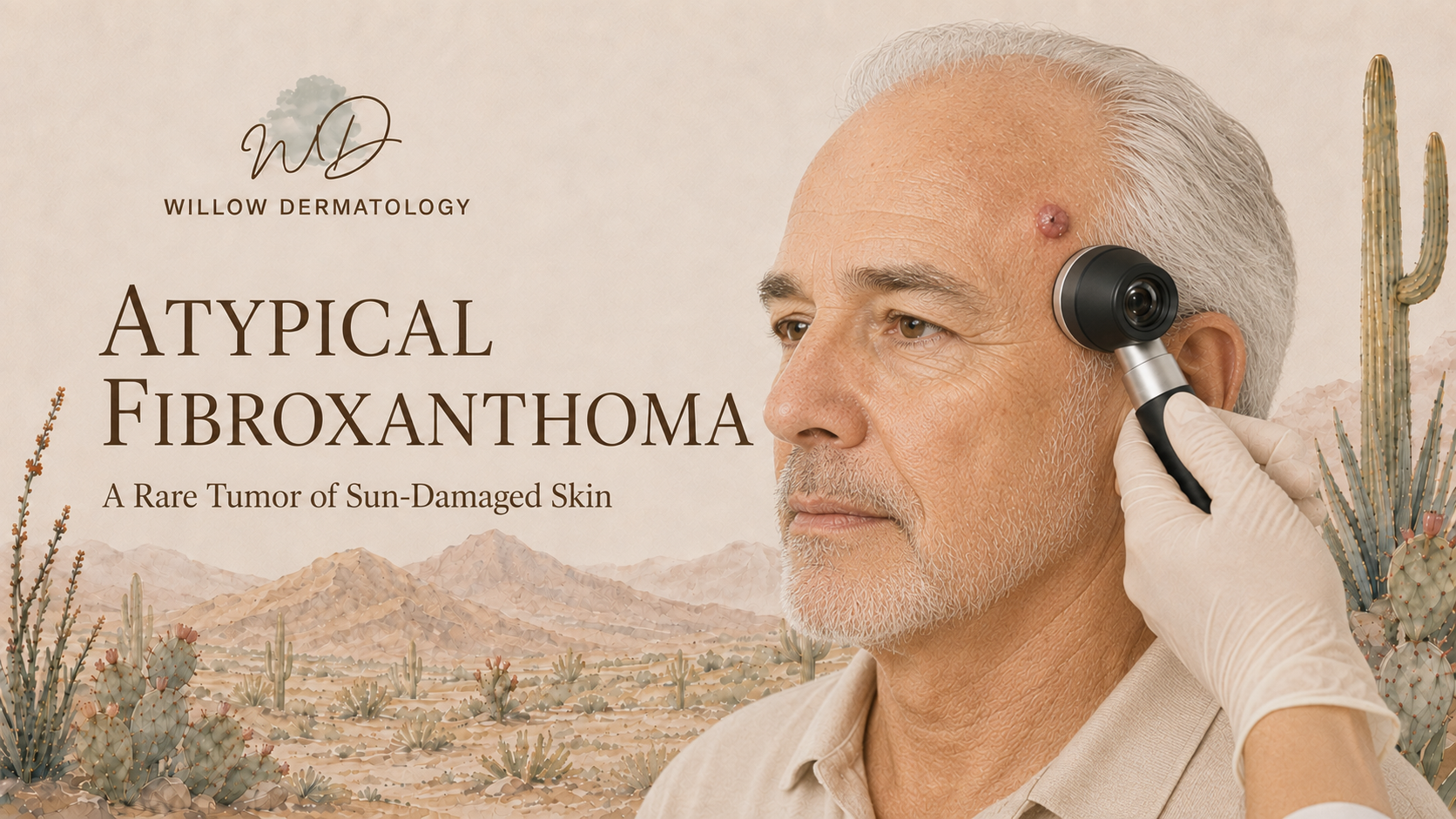
Atypical fibroxanthoma is a rare superficial tumor that usually develops on chronically sun-damaged skin in older adults. It most often occurs on the scalp, face, ears, or neck and may grow rapidly over several weeks or months.
The tumor commonly appears as a firm pink or red dome-shaped nodule. It may develop scale, crusting, tenderness, or occasional bleeding and can resemble squamous cell carcinoma or another common skin cancer.
Diagnosis requires a biopsy and specialized laboratory stains. A sufficiently deep specimen is important to determine whether the tumor is confined to the superficial skin or has features of a more aggressive related cancer called pleomorphic dermal sarcoma.
Mohs micrographic surgery or surgical excision with careful margin assessment is commonly used to remove atypical fibroxanthoma. Most superficial tumors have a favorable outcome after complete removal, but continued surveillance is recommended because recurrence and rare spread can occur, particularly in immunosuppressed patients or tumors with higher-risk features.
Cutaneous Metastasis of Internal Malignancy
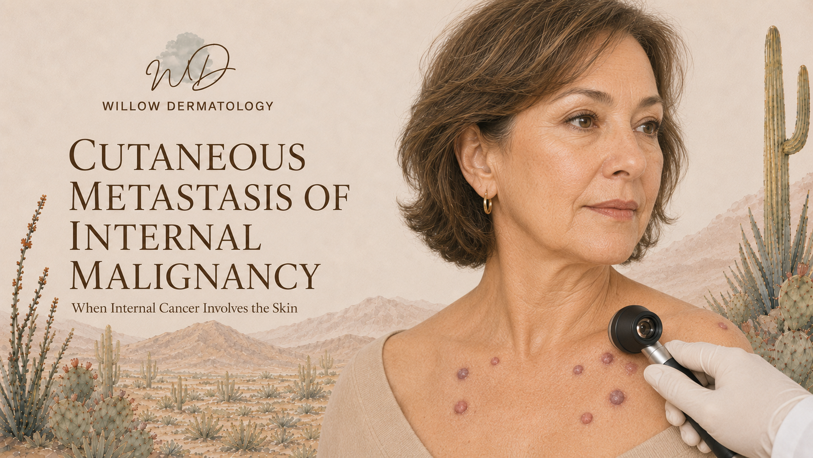
A cutaneous metastasis occurs when cancer cells from an internal organ spread to the skin. This may develop in someone with a known history of cancer, signal that a previous cancer has returned, or occasionally be the first visible sign of an undiagnosed internal malignancy.
Cutaneous metastases most often appear as one or more firm, painless, rapidly developing nodules. They may be skin-colored, pink, red, brown, or violet. Depending on the original cancer, lesions may occur on the scalp, chest, abdomen, back, or near a previous surgical scar.
Diagnosis requires a skin biopsy. Specialized immunohistochemical and molecular testing may help determine where the cancer originated. Additional imaging, laboratory testing, and review of previous pathology are often necessary.
Treatment is directed by the type and extent of the underlying internal cancer. Systemic therapy may include immunotherapy, targeted therapy, hormonal therapy, or chemotherapy. Surgery or radiation therapy may be used for selected isolated lesions or to relieve discomfort, bleeding, or other symptoms. Care is coordinated closely with medical oncology and other appropriate specialists.
Extramammary Paget Disease
Extramammary Paget disease is a rare cancer that begins within the outermost layer of skin in areas containing apocrine glands. It most commonly affects the vulvar, groin, perianal, scrotal, penile, or underarm skin.
It often appears as a persistent red, pink, or discolored scaly plaque. Symptoms may include itching, burning, tenderness, irritation, or bleeding. Because it can resemble eczema, a fungal infection, or contact dermatitis, diagnosis is frequently delayed when a rash does not improve with routine treatment.
A skin biopsy is required for diagnosis. Multiple biopsies or mapping biopsies may be needed because cancer cells can extend beyond the visible borders of the rash. The pathology examination also determines whether the cancer remains within the outer layer of skin or has become invasive.
Extramammary Paget disease may begin primarily in the skin or represent spread from an underlying genitourinary or gastrointestinal cancer. Additional screening is individualized according to the tumor location, pathology findings, symptoms, age, and medical history.
Mohs micrographic surgery, staged margin-controlled excision, or wide local excision may be used to remove the cancer. Radiation therapy or other localized treatments may be considered for selected noninvasive tumors, recurrent disease, or patients who are not surgical candidates. Invasive or metastatic disease may require lymph node evaluation and coordinated oncology care.
Long-term surveillance is important because extramammary Paget disease can recur even after apparently complete treatment.
When to Schedule an Evaluation
Rare skin cancers can closely resemble benign or inflammatory conditions. Schedule a dermatologic evaluation for:
A firm growth that is steadily or rapidly enlarging
A persistent eyelid nodule or inflammation that does not improve
A new growth developing within a scar or previously treated area
Multiple unexplained skin nodules
A persistent groin, genital, perianal, or underarm rash that does not respond to treatment
A lesion associated with numbness, tingling, pain, bleeding, or loss of normal function
Early biopsy and accurate diagnosis allow treatment to begin before a rare skin cancer becomes more extensive.